Innovative Method of Local Anesthesia for Penile Surgery
introduction

To help alleviate our patients’ anxiety and physical discomfort during surgery, we routinely apply acupuncture as an adjuvant to local anesthesia (no more than 10% males require an intravenous sedation); we find it to be consistently effective in this regard, although we acknowledge that neither acupuncture’s mechanism nor its role as an anesthetic or analgesic is without controversy.1 We regularly focus on the following acupoints (dots, Figure 1): Hegu (LI 4), Shou San Li (LI 10), Quchi (LI 11), Neiguan (PE 6) and Shen Men (H 7). The Hegu acupoint is positioned at the highest point where the thumb and the index finger adduct; the Quchi point at the lateral end of the “transverse cubital crease,” with the elbow flexed at a right angle; and the Shou San Li point a 3-finger breadth caudally to the Quchi acupoint.
The Neiguan is positioned 5 cm proximal to the middle point of the volar transverse carpal crease, between the flexor carpi radialis muscle and the palmaris longus tendon, with the forearm supine. The Shen Men is at the cross of the wrist proximal transverse crease and the low-lateral margin of the pisiform bone. This innovative, acupuncture-assisted local anesthesia was first implemented in male potency reconstructive strategies in 1988. It has been modified to ensure that surgical patients may be discharged on an ambulatory basis; acupoints may be further updated for the physician's convenience or at the patient's discretion. Although it is not universally accepted as valid, it is worth noting that acupuncture was included in UNESCO's lists of intangible cultural heritage on November 16, 2010. Further research is warranted.
Methods of local anesthesia
Over the last few decades, several types of nerve blockades2,3,4,5,6 have been recommended for penile surgeries with the patients under local anesthesia rather than the well-established general or spinal anesthesia. However, either an inconvenient and unpleasant pudendal nerve block, or an adjuvant intravenous injection of sedatives, has been unavoidable.7,8,9,10 We would like to describe local anesthetic methods for penile surgeries on an outpatient which include proximal dorsal nerve block,11,12 peripenile infiltration, penile crural block,13 cavernous nerve blockade, and topical injection of the medial low abdominal region as necessary. We believe that these local anesthetic techniques offer an optimal alternative to the methods typically used in various penile surgeries.
The innervations of the human penis
The human penis is a unique structure composed of multiple fascial layers which surround the three cylinders of ereinnervations tile sinusoids, the most ideal milieu in the human body in which to apply Pascal's law.14 Thus, it consists of the glans penis, the corpus spongiosum with the bulb of the urethra, the paired corpora cavernosa and the bulbospongiosus, as well as the ischiocavernosus muscles. It is the most sensitized coman in the human body owing to its dense nervous distribution.15 A full review of the nervous pathway and neurophysiology16 of the human penis is not necessary to administer local anesthesia. However, one must always keep in mind the relationship between the cavernous nerve and the pudendal nerve to the bony and fibromuscular structures of the penis, in order for the surgeon to be able to reproduce these nervous blockades. At the level of penile hilum, where the distal third urethral bulb is encountered and the penile crura are formed, the fibers of the cavernous nerve are located at around the medial third of each corpus cavernosum. Some of these cavernous fibers enter the corpora cavernosa and corpus spongiosum laterally, thus abutting with the cavernous and urethral arteries. The remaining fibers travel distally with the dorsal nerve and enter the corpus cavernosum as well as the corpus spongiosum in various sites to innervate the mid and distal penile shaft. Meanwhile, the paired pudendal nerves, with somatomotor as well as somatosensory components, are located a finger’s breadth cranio-posteriorly and are regarded as the starting point of the proximal dorsal nerve. The somatic sensory nerves originate at the receptors in the penile skin and glans penis. Subsequently, signals carrying sensory information of pain and temperature ascend via the spinothalamic tract; while vibratory stimuli are carried in the dorsal column; and touch and pressure sensations are transmitted via both pathways to the thalamus. All the transmitting signals of the motor nerve are sent from lateral to medial tracts, and vice versa for the sensory component. Therefore, a lateral blockade of a certain nerve can always result in an anesthetic effect to its medial innervating proper. It is a rule that the anatomical arrangement of the deep dorsal vein, dorsal artery and dorsal nerve is arrayed from the medial side. Although a thorough understanding of the penile neuro-anatomy is indispensable for a well-performed penile implantation, extensive knowledge of the fibro-muscular skeleton of the human penis is also a prerequisite and is clinically meaningful, since through recognition the bony or fibro-muscular landmarks helps to precisely localize any specific nerve.
The fibro-muscular skeleton of the human penis (Fig. 2)
The traditional anatomical description of the human penis has been well-established. However, it may not sufficiently detail a precise fibrous landmark, the knowledge of which is essential to induce adequate nerve blockade during outpatient penile surgeries. Fortunately, recent studies have reported more knowledge about the penile anatomy, which will benefit the surgeon in recognizing the detailed fibrous landmarks within the penis. This includes the tunica albuginea,17 the exact position and relationship between the skeletal muscle and smooth muscles,18 and the upper border as well as the lower margin of the symphysis pubis and the ischial tuberosity. The tunica albuginea is consistently described as a single layer with uniform thickness and strength circumferentially.19 It is, indeed, a bi-layered structure which can be divided into an inner circular layer and an outer longitudinal layer. Its thickness as well as its strength can vary significantly depending on its specific position. The outer longitudinal layer is an incomplete coat which is absent between the 5 and 7 o'clock positions, where two triangular ligamentous structures form. These structures, termed the ventral thickening (VT), are a continuation of the anterior fibers of the left and right bulbospongiosus muscles, respectively. A weak border is positioned ventrally between two ventral thickenings, and a hazardous prosthesis extrusion could potentially occur in this region.20 On the dorsal aspect, between the 1 and 11 o'clock positions, is the region called the dorsal thickening, a radiating aspect of the bilateral ischiocavernosus muscles. It then converges distally and is arranged centrally to form a distal ligament (DL), which is located immediately above the 12 o'clock position of the navicular fossa of the distal urethra and acts as a trunk of the glans penis.21,22 Without this strong ligament, the glans would be too weak to bear the buckling pressure generated during coitus. A surgeon should be able to feel five prerequisite fibrous or bony landmarks of the penis prior to performing nervous blockage. First of all, the distal tip of the distal ligament can be clearly perceived if one puts the finger palm of one's index finger, for example, over the glanular tip. Second, the borders of the corpus spongiosum formed by bilateral ventral thickenings can be felt when one puts his or her finger palm over the penoscrotal junction of the patient. This is more easily palpable if the patient constricts his anus. Third, one is able to tell whether the patient's penile hilum is fibrotic or not by feeling whether the underlying suspensory ligament is free of resistance—to do so, one uses a gentle pushing force along the pubic angle or from the lateral aspect when the penile shaft is pulled away from the body axis. When the hilum is less fibrotic, it is easier to introduce the injection needle, thus allowing for a more precise proximal dorsal nerve block. Fourth, one has to be sure of the exact discrimination between the borders of the upper and lower margins of the symphysis pubis. Finally, the ability to palpate the ischial tuberosity is required.
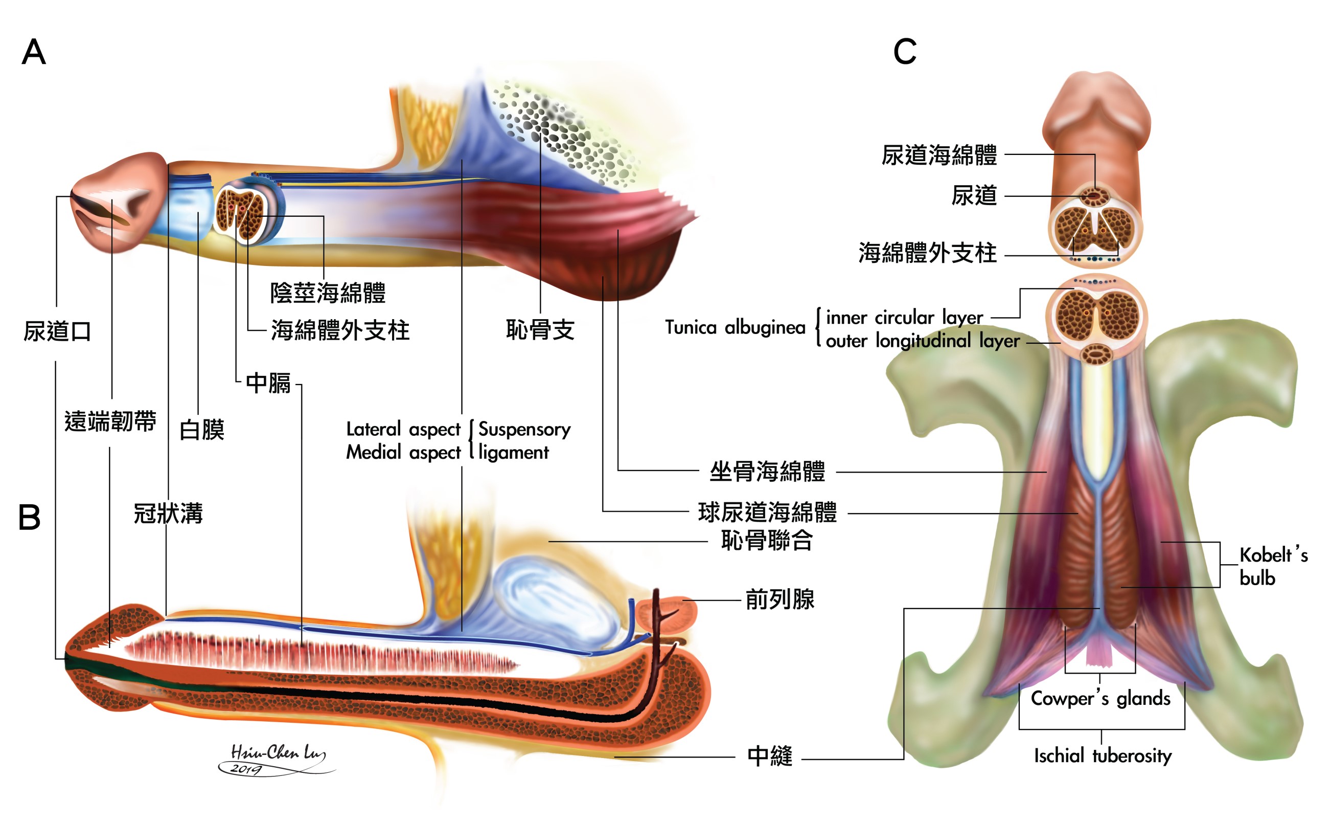
The corpora cavernosa are surrounded by the tunica albuginea, which, again, is a bi-layered structure with an inner circular and an outer longitudinal layer with multiple sub-layers. The incomplete septum is dorsally fenestrated. The intra-cavernosal pillars, which may be considerably larger distally, are a continuation of the inner circular layer. The distal ligament is aggregated from the collagen bundles of the outer longitudinal layer of the tunica albuginea. It is an inelastic fibrous structure which forms the trunk of the glans penis. The ischiocavernosus muscle is paired and situated at the lateral boundary of the perineum. Each segment covers its ipsilateral penile crus. Meanwhile, the anterior fibers of the bulbospongiosus muscle partially spread out to encircle the corpus cavernosum and mostly insert into the ventral thickening of the tunica.
Proximal dorsal nerve block and Peripenile infiltration (Figure 3)
A 23-gauge, 1.25' (3.18-cm) disposable needle connected to a 10-mL syringe was used to inject the local anesthetic, a 0.8% Lidocaine solution prepared in an aseptic bowl pre-filled with 1.0 mL of a 1:200,000 epinephrine solution. With the bevel parallel to the longitudinal body axis, the needle is introduced in-between the suspensory ligaments along the pubic angle while the penile shaft is pulled a little caudally away from the body axis by the surgeon's left hand. Then the injection is made in three directions in order to cover the proximal dorsal nerves bilaterally. An aspiration of the syringe is made before any attempt at injection in order to avoid inadvertent entry into the vessels. Under a finger guide, the needle is withdrawn just enough to free it from being entrapped in the penile hilum. The needle is then shifted laterally and advanced to the lateral margin of the penile crus; then an injection is slowly delivered while the needle is withdrawn until the sub-cutaneous space is encountered. The needle is advanced caudally and further infiltration is made after ensuring no inadvertent entry into a vessel. The contralateral side is anesthetized in a similar fashion.
Two underlying rigid borders are felt by palpation while pushing downward from the penoscrotal junction. A right-handed surgeon requires the patient's glans penis to be held upward by an assistant's left hand with the palm of the index finger and thumb pinch the 3 and 9 o'clock positions respectively at the retrocoronal sulcus. Then, a rapid and precise puncture is made at the intersection of the medial raphe and the penoscrotal junction. Subsequently, a meticulous injection of the ventral thickening is made bilaterally from its medial margin. The peripenile infiltration is performed in a semi-circumferential manner unilaterally, and then the infiltration of the contralateral side is made in a similar fashion to complete the circle in the ventral aspect. Thus, topical infiltration of the junction between the corpus spongiosum and the corpora cavernosa is mandatory in order to avoid an incomplete block of the sensitive corpus spongiosum. Care should be taken not to puncture the paper-thin tunica albuginea of the corpus spongiosum. A gauge compression of the bleeding point for several minutes is sufficient to stop the bleeders if the spongiosal body is incidentally entered. The scrotal infiltration may be extended caudally if a water reservoir is intended to implant in the scrotal pouch or a 90-degree Z-plasty is intentionally performed. Finger-guided manipulation is very helpful for the entire procedure.
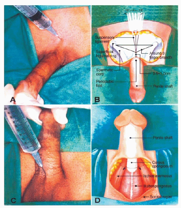
The patient is put in the supine position which is suitable for the entire operation
- Proximal dorsal nerve block, the needle, with its bevel parallel to the direction of the body axis, was quickly inserted 0.5 to 1.0 cm cranial to the penopubic fold in between the suspensory ligament until the infrapubic angle was encountered.
- Illustration of the precise positions injected: It was injected in three directions, medially and 15-degree obliquely bilaterally, in the penile hilum in order to cover the bilateral dorsal nerves.
- Ventral infiltration: The peripenile space is meticulously infiltrated one and half finger-breadths below the penoscrotal junction while a finger-guided manipulation (utilizing the index finger of the assistant's hands) is used to confirm the precise position of the injection.
- Illustration of its anatomical landmarks. The peripenile injection shall be made to encircle the penile shaft. Note that the junction between the corpus spongiosum and the corpora cavernosa (arrow), i.e. the 5 and 7 o'clock positions shall be exactly blocked; otherwise, an incomplete blockage will be unavoidable. The needle is subsequently withdrawn and advanced laterally to inject the lateral aspect of each corresponding crus. (This photo was reproduced by courtesy of the Journal of Andrology.)
With the bevel parallel to the longitudinal body axis the needle is introduced in-between the suspensory ligaments along the pubic angle while the penile shaft is pulled a little caudally away from the body axis by the surgeon's left hand. Then the injection is made in three directions in order to cover the proximal dorsal nerves bilaterally. An aspiration of the syringe is made before any attempt of injection in order to avoid inadvertent entry into the vessels. Under a finger guide, the needle is withdrawn back just sufficiently to free it from being entrapped in the penile hilum. The needle is then shifted laterally and advanced to the lateral margin of penile crus; then an injection is slowly delivered while the needle is withdrawn until the subcutaneous space is encountered. The needle is advanced caudally and further infiltration is made after ensuring no inadvertent entry into a vessel. The contralateral side is anesthetized in a similar manner.
The glans penis is to be held upward by an assistant's left hand with the palm of the index finger and thumb pinching the 3 and 9 o'clock positions respectively at the retrocoronal sulcus. Then a rapid and precise puncture is made at the intersection of the medial raphe and the penoscrotal junction. Subsequently, a meticulous injection of the ventral thickening is made bilaterally from its medial margin. The peripenile infiltration is performed in a semi-circumferential manner unilaterally, and then the infiltration of the contralateral side is made in a similar fashion to complete the circle in the ventral aspect.
Penile crural block and Cavernous nerve blockage (Figure 4)
The penile shaft is put in a pendulous position while the patient is in a comfortable supine position. A 23G x 1.5' (3.81-cm)-long disposable needle is punctured into the skin at the intersection of the penopubic fold, one finger’s breadth laterally. Under finger guidance, the needle is pushed downward vertically along the pubic angle until the medial third penile crus is targeted. It is then withdrawn a little upward before the local anesthetic solution is delivered in case of inadvertent puncture into the corpus. A bloody aspiration signals entry into the corpus cavernosum. An experienced hand can feel whether an inadvertent puncture through the tunica has been made since the tunica can act as a barrier in providing an intermediate resistance. An injection of a 2 to 3 mL solution is sufficient to block the neurofibers of the cavernous nerve. Under finger guidance, the needle is withdrawn sufficiently to free it from being entrapped in the penile hilum. The needle is then advanced to the lateral margin down to the ischial tuberosity. A slow and even delivery of the local anesthetic solution is made while the needle is withdrawn superficially until the subcutaneous space is met.
A 23-gauge, 1.5' (3.81-cm) disposable needle is recommended for this purpose. The penile shaft is stretched upward while the penoscrotal junction is identified. The needle is targeted at a 45° angle oblique to the coronal plane at the junction of the corpus spongiosum and the penile crus. It is advanced to about 2 cm in order to block the cavernous nerve. Thus, there are two methods for performing the cavernous nerve blockade, dorsally and ventrally. An additional topical anesthetic injection is required if implantation of a three-piece prosthesis is performed. Similarly an extended anesthesia of the involved tissues is mandatory whenever surgery requires.
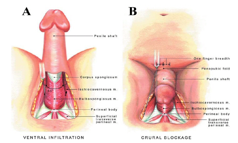
The patient is put in the supine position which is suitable for the entire operation
- Ventral infiltration: After proximal dorsal nerve blockage is performed, the peripenile space is meticulously infiltrated one and half finger-breadths below the penoscrotal junction. Then the needle is targeted 45 degrees oblique to the coronal plane and inserted deeper in order to block the cavernous nerve.
- Crural blockage: the needle is quickly inserted into the subcutaneous space at the point crossing the penopubic fold and one finger-breadth bilaterally, respectively. Then it is depressed to target one-third of medial crus in order to block the cavernous nerve. Subsequently the needle is withdrawn and advanced laterally to inject the lateral aspect of each corresponding crus. (This photo is reproduced by courtesy of the International Journal of Andrology.)
The penile shaft is put in a pendulous position while a 23G x 1.5' (3.81-cm)-long disposable needle is punctured into the skin at the intersection of the penopubic fold one finger-breadth laterally. Under finger guidance, the needle is pushed downward vertically along the pubic angle until the medial third penile crus is targeted. Under finger guidance the needle is then withdrawn sufficiently to free it from being entrapped in the penile hilum. The needle is then advanced to the lateral margin down to the ischial tuberosity. A slow and even delivery of the local anesthetic solution is made while the needle is withdrawn superficially until the subcutaneous space is met and then advanced laterally to inject the lateral aspect of each corresponding crus.
Local anesthesia performed on an outpatient basis for penile surgeries appears to be highly promising.23-27 Our study shows that local anesthetic methods of proximal dorsal nerve block, peripenile infiltration, penile crural block, cavernous nerve blockage, and a topical injection have, on an outpatient basis, been proven to be reliable, simple, and safe, with few complications in our study. Local anesthesia offers the advantages of fewer anesthetic adverse effects, reduced morbidity, greater protection of the patient's privacy, and a more rapid return to daily activity with minimal complications. Its success, though, depends on a pre-requisite knowledge of penile anatomy; to this end, we offer a number of publications.28-31 We hope that this practical method can be shared and disseminated in medical communities across the world.
References
- Hsu GL.: Peyronie’s disease. In: APSIR BOOK on Erectile Dysfunction, 1st ed. Edited by Kim, Y. C. and Tan, H. M. Malaysia: Pacific Cosmos Sdn Bhd, chapt. 18, pp. 200-212, 1999.
- Kirya C, Werthmann MW Jr. Neonatal circumcision and penile dorsal nerve block--a painless procedure. J Pediatr. 1978; 92: 998-1000.
- Brown TC, Weidner NJ, Bouwmeester J. Dorsal nerve of penis block- anatomical and radiological studies. Anaesth Intensive Care. 1989; 17: 34-38.
- Dunn RL, Harris DL. Technique for continuous dorsal penile nerve anaesthesia following penile surgery. Br J Surg. 1997; 84: 220-221.
- Ghanem H, Fouad G. Penile prosthesis surgery under local penile block anesthesia via the infrapubic space. Int J Androl. 2000; 23: 357-359.
- Dos Reis JM, Glina S, Da Silva MF, Furlan V. Penile prosthesis surgery with the patient under local regional anesthesia. J Urol. 1993; 150: 1179-1181.
- Kaufman JJ. Penile prosthesis surgery under local anesthesia. J Urol. 1982; 128: 1190-1191.
- Light JK, Scott FB. Implantation of the inflatable penile prosthesis using local anesthesia. In: Kaye KW, d. Outpatient Urologic Surgery. Philadelphia: Lea & Febiger; 1985; pp. 261-268.
- Scott FB. Outpatient implantation of penile prostheses under local anesthesia. Urol Clin North Am. 1987; 14: 177-185.
- Leach GE. Local anesthesia for urological procedures. Urology. 1996; 48; 284-288.
- Hsu GL. Chen SH. Weng SS. Out-patient surgery for the correction of penile curvature. Br J Urol. 1997; 79: 36-9. (Correspondent and principal author)
- Hsu GL. Hsieh CH. Wen HS. Hsieh JT and Chiang HS: Outpatient surgery for penile venous patch with the patient under local anesthesia. J Androl. 2003; 24: 35-39. (Correspondent and Principal author)
- Hsu GL. Hsieh CH. Wen HS, Chen SC, Chen YC, Liu LJ, Mok MS and Wu CH. Outpatient penile implantation with the patient under a novel method of crural block. Int J Androl. 2004; 27: 147-151. (Correspondent and Principal author)
- Hsu GL: The hypothesis of human penile anatomy, erection hemodynamic and their clinical applications. Asian J Androl.2006; 8: 225-234,. (Invited)
- Breza J, Aboseif SR, Orvis BR, Lue TF, Tanagho EA. Detailed anatomy of penile neurovascular structures: surgical significance. J Urol. 1989; 141: 437-443.
- Salmons S. Muscle: Muscles and Fasciae of the Trunk. In: Bannister LH, Berry MM, Collins P, Dyson M, Dussek JE, Ferguson MWJ, editors. Gray's Anatomy. London: Churchill Livingstone; 1995. pp. 819-29.
- Hsu GL, Brock G, Martinez-pinerio L, et al: The three-dimensional structure of the human tunica albuginea: Anatomical and Ultrastructural Level. Int J Impot Res. 1992; 4: 117-29.
- Hsu GL. Hsieh CH. Wen HS, Hsu WL and Chen CW: Anatomy of the human penis: The relationship of the architecture between skeletal and smooth muscles. J Androl. 2004; 25: 426-431. (Correspondent and Principal author)
- Eardley I, Sethia K, Anatomy and Physiology of Erection. In: Eardley I., Sethia K., Editors. Erectile Dysfunction, Current Investigation and Management. London: Mosby; 2003. pp. 7-23.
- Hsu GL, Brock G, Martinez-Pineiro L, von Heyden B, Lue TF, Tanagho EA. Anatomy and strength of the tunica albuginea: its relevance to penile prosthesis extrusion. J Urol. 1994; 151: 1205-8.
- Hsu GL, Lin CW, Hsieh CH, Hsieh JT, Chen SC, Kuo TF, Ling PY, Huang HM, Wang CJ, Tseng GF. Distal ligament in human glans: a comparative study of penile architecture.J Androl. 2005; 26: 624-628,. (Correspondent and Principal author)
- Hsu GL, Chen HS, Huang SJ. Does tunica anatomy matter in penile implant? Transl Androl Urol. 2014; 4: 406-412. doi: .3978/j.issn.2223-4683.2014.03.04 (Correspondent and Principal author)
- Hsu GL, Hsieh CH, Chen HS, Ling PY, Wen HS, Huang HM, Liu LJ, Chen CW, and Chua c. The Advancement of Pure Local Anesthesia for Penile Surgeries: Can an Outpatient Basis be Sustainable? J Androl. 2007; 28: 200-205. (Correspondent and Principal author)
- Hsieh CH, Liu SP, Hsu GL Chen HS, Molodysky E, Chen YH, Yu HJ. Advances in our understanding of mammalian penile evolution, human penile anatomy and human erection physiology: Clinical implications for physicians and surgeons. Med Sci Monit. 2012; 18: RA118-125. (Correspondent and Principal author).
- Hsu GL. “Physiological approach to penile venous stripping surgical procedure for patients with erectile dysfunction (Patent No.: US 8,240,313, B2). “http://www.google.com/patents/US20110271966.
- Hsu GL, Molodysky E, Liu SP, Hsieh CH, Chen HC, Chen YH. A Combination of Penile Venous Stripping, Tunical Surgery and Varicocelectomy for Patients with Erectile Dysfunction, Penile Dysmorphology and Varicocele under Acupuncture-aided Local Anesthesia on Ambulatory Basis. Surgery: current research. 2013; S12: 008. doi:10.4172/2161-1076.S12-008 (Correspondent and Principal author)
- Hsu GL, Zaid UX, Hsieh CH, Huang SJ. Acupuncture assisted regional anesthesia for penile surgeries. Transl Androl Urol. 2013; 2: 291-300. doi: 10.3978/ j.issn.2223-4683.2013.12.02 (Invited, correspondent and Principal author)
- Hsu, G-L. (2018). Erection Abnormality. In M. K. Skinner (Ed.), Encyclopedia of Reproduction. vol. 1, pp. 382–390. Academic Press: Elsevier. http://dx.doi.com/10.1016/B978-0-12-801238-3.64374-X
- Hsu, G-L., & Liu, S-P. (2018). Penis Structure. In M. K. Skinner (Ed.), Encyclopedia of Reproduction. vol.1, pp. 357–366. Academic Press: Elsevier. http://dx.doi.com/10.1016/B978-0-12-801238-3.64602-0
- Hsu, G-L., & Lu, H-C. (2018). Penis Structure—Erection. In M. K. Skinner (Ed.), Encyclopedia of Reproduction. vol. 1, pp. 367–375. Academic Press: Elsevier. < href="http://dx.doi.com/10.1016/B978-0-12-801238-3.64603-2" target="_blank">http://dx.doi.com/10.1016/B978-0-12-801238-3.64603-2
- Huang, P-C., & Hsu, G-L. (2018). Vascular Surgery for Erectile Dysfunction. In M. K. Skinner (Ed.), Encyclopedia of Reproduction. vol. 4, pp. 427–436. Academic Press: Elsevier. http://dx.doi.com/10.1016/B978-0-12-801238-3.64804-3
 Images on this webpage are licensed under a CC Attribution-ShareAlike 4.0 International License.